Institute of Biotechnology - Life Sciences Centre - Vilnius University

Research areas
Biophysical studies
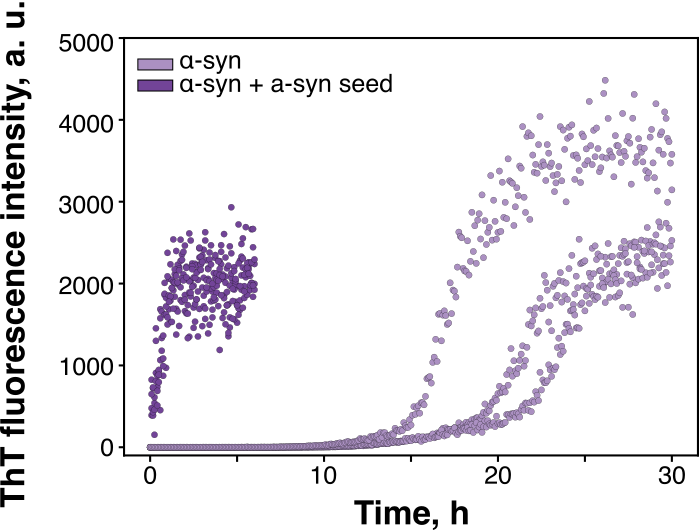
Many proteins can switch from their normal, functional state into amyloid fibrils – self-propagating aggregates linked to Parkinson’s, Alzheimer’s and other diseases. We investigate how these fibrils form and propagate, and how liquid–liquid phase separation, where proteins condense into liquid-like droplets, influences this process.
Using amyloid-specific fluorescent dyes, mass spectroscopy, aggregation kinetics and microscopy, we follow aggregation from the first oligomers to mature fibrils. We focus on how conditions inside protein droplets affect which fibril structures form, including in α-synuclein and the 14-3-3 protein family
Key publications:
Structural biology
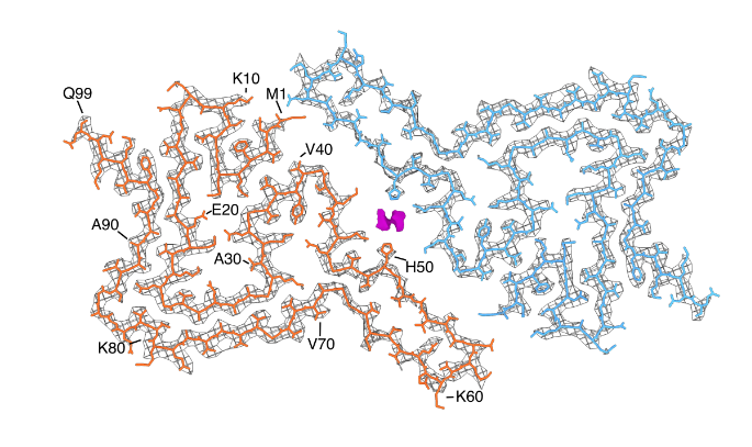
The same protein can assemble into amyloid fibrils with different structures – so-called strains – and these differences can change how fibrils grow, spread and affect cells. To understand the mechanisms behind this, we study fibrils at the molecular level.
We combine circular dichroism (CD) and Fourier-transform infrared spectroscopy (FTIR) to follow changes in secondary structure, atomic force microscopy (AFM) to see fibril shape, and cryo-electron microscopy (cryo-EM) to resolve fibril structures in detail. This lets us link the conditions in which fibrils form to the structures they end up with.
Key publications:
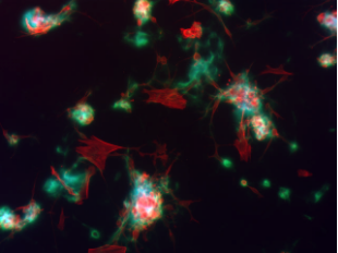
Cellular studies
Understanding how proteins interact in a test tube is only part of the picture. To see how amyloid aggregates behave in a biological context, we also study their effects on cells.
We measure cell viability with the MTT assay, which reports on cells’ metabolic activity, and the LDH assay, which detects damage to the cell membrane. This lets us compare how toxic different aggregates and fibril types are, and test whether potential inhibitors reduce that harm
Key publications:
- Formation of amyloid fibrils by the regulatory 14-3-3ζ protein, Open Biology
- Imidazo[2,1‑b][1,3]thiazine Derivatives as Potential Modulators of Alpha-Synuclein Amyloid Aggregation, ACS Chemical Neuroscience